


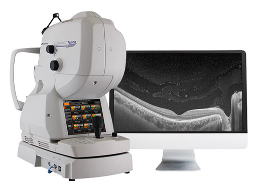
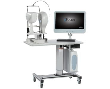
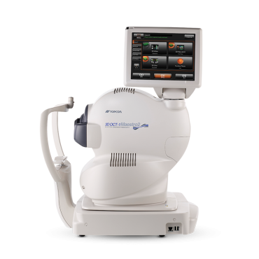
Topcon DRI OCT Triton Swept Source OCT
Brand: Topcon
SKU: OT0TO39031KTFG
Price:
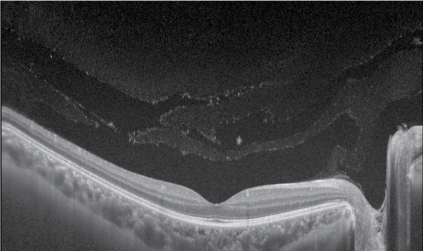
The Topcon DRI OCT Triton Swept Source OCT uses swept-source technology to allow visualization into the deepest layers of the eye – even through cataracts, hemorrhages, gas bubbles and other media opacities, making it possible for more patients to be imaged.
Free
MSRP:
The fast 100 kHz scanning speed and invisible scan beam rapidly capture detailed images, resulting in fewer motion artifacts and stunning image quality. Decrease chair time and improve your clinical workflow with fewer rescans and multimodal imaging including, OCT, true-color fundus photography, FA1 and FAF1.
FEATURES:
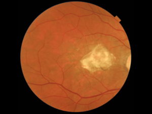
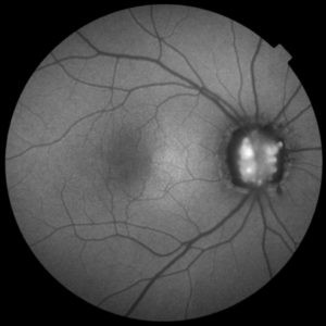
- HIGH RESOLUTION: Multimodal platform provides easy, yet comprehensive comparison of microvascular impairment with FA1, FAF1, OCT and color fundus images.
- SCAN MORE PATIENTS: Swept-source technology allows imaging through media opacities
- FEWER RESCANS: Invisible scan beam allows patients to focus on the fixation target and reduce involuntary eye movement
- WIDE SCAN: A 12mm x 9mm scan encompasses the optic nerve and macula and can be acquired in 1.8 seconds for fast assessment of the posterior pole.
- RICH, DETAILED IMAGES: Image quality is further enhanced by PixelSmart® Technology2
1DRI OCT Triton Plus only.
2PixelSmart® is a function of IMAGEnet 6 software.
SPECIFICATIONS
Fundus Imaging
- Imaging Modes Color, FA,* FAF,* Red-Free,** IR
- Field of View 45° / 30° (Digital Zoom)
- Operating Distance 34.8mm
- Minimum Pupil Diameter Ø4.0mm / Small Pupil Mode: Ø3.3mm
- Resolution (On Fundus)
- Center: 60 Lines/mm or more,
- Middle (r/2): 40 Lines/mm or more,
- Periphery (r): 25 Lines/mm or more
OCT
- Scan Range (On Fundus) 6 to 12mm
- Scan Patterns
- 3D Wide: 12x9mm
- 3D Macula: 7x7mm
- 3D Optic Disc: 6x6mm
- Combination Scan: 12x9mm + 5 Line Cross
- Line: 6-12mm
- 5 Line Cross: 6-12mm
- Scan Speed 100,000 A-Scans Per Second
- Lateral Resolution 20 μm
- Axial Resolution
- Optical: 8 μm
- Digital: 2.6 μm
- Minimum Pupil Diameter Ø2.5mm
- Fixation Target Internal Fixation Target/ Peripheral Fixation Target / External Fixation Target
- Diopter Range
- Without the diopter compensation lens: -13D to +12D
- When the concave compensation lens is used: -12D to -33D
- When the convex compensation lens is used: +11D to +40D
Anterior Segment***
- Photography Type IR
- Operating Distance 17mm
- Scan Range (On Cornea) 3 to 16mm
- Scan Patterns Line Anterior Segment: 3-16mm / Radial Anterior Segment: 6-16mm
- Fixation Target Internal Fixation Target / External Fixation Target
* FA photography and FAF photography can be performed in only DRI OCT Triton (plus).
** Digital red-free
*** Observation & photography of anterior segment can be performed only when the anterior segment attachment kit is used.
Related Products
A Division of Advancing Eyecare™
© 2024 Lombart Healthcare All Rights Reserved.
Account Information
Get to Know Lombart
Lombart Healthcare is committed to keeping our site accessible to everyone. We welcome feedback on ways to improve the site’s accessibility so it is easy for everyone to navigate.
Free shipping is only available for online orders in the continental United States.
Certain exclusions apply.
Exclusions include, but are not limited, to the following products:
Acuity Systems & Projectors, Chair & Stand Accessories, Autorefractors, Lensmeters, Keratometers, Portable Slit Lamps, Stools, Tables, Tonometers, Trial Lens Sets.
- Exam Lane
- Pre-Test
- Diagnostic/Imaging
- Treatment & Surgical
- Lab & Dispensing
- Lens Edgers
- Supplies & Accessories
- Pre-Owned
- All Pre-Owned Equipment
- Pre-Owned Autorefractor/Keratometers
- Pre-Owned Biometers
- Pre-Owned BIOs
- Pre-Owned Chairs & Stands
- Pre-Owned Edgers
- Pre-Owned Lasers
- Pre-Owned Lensmeters
- Pre-Owned Manual Keratometers
- Pre-Owned OCTs
- Pre-Owned Projectors
- Pre-Owned Refractors & Phoroptors
- Pre-Owned Retinal Cameras
- Pre-Owned Slit Lamps
- Pre-Owned Tonometers
- Pre-Owned Ultrasounds
- Pre-Owned Visual Field Perimeters